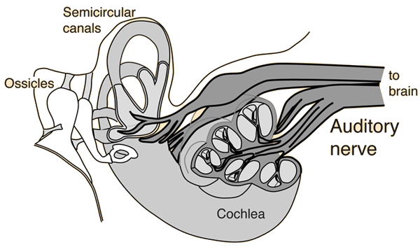

This segregation of high and low frequencies persists throughout the CNS. Low frequency fibers then pass in the central core of the VIIth nerve surrounded by high frequency fibers (see Auditory System: Structure and Function).
AUDITORY NERVE PATHWAY CODE
Click on the cochlea in Figure 13.3 to see the color code of pitch, as if the cochlea were a piano. This starts with high frequencies transduced at the base of the cochlea, and low frequencies transduced at the apex (see Figure 12.7). Afferents from this longitudinal strip on the superior temporal gyrus diverge to a wide variety of other cortical processing areas, including Wernicke’s area in the parietal lobe where speech is processed.Īuditory afferents are tonotopically organized from the ear to the cortex. Primary auditory cortex, or Herschel’s gyrus in insular cortex, is tonotopically organized. The two different patterns of dashed lines combine to form a solid line above the superior olive, meant to indicate the combination of monaural inputs into bilateral and binaural activation. Then, click on the cochlea and text to gain further information.įigure 13.3 shows the same detail processing system as in Figure 13.2, only now with the more realistic situation of input from both ears. Thalamic afferents reach the superior temporal gyrus through the sub-lenticular portion of the internal capsule.īinaural auditory afferents.

All afferents then synapse in the medial geniculate body of the thalamus. Most of these afferents synapse in the inferior colliculus. Some nuclei within the lateral lemniscus further process the sound. The output of the superior olive travels in the lateral lemniscus. Thus, cells in the superior olive receive inputs from both ears and are the first place in the central auditory system where binaural processing (stereo hearing) is possible. Some fibers from the ventral cochlear nucleus cross the midline in the trapezoid body. Fibers from the ventral cochlear nucleus synapse in the ipsilateral and contralateral superior olivary nucleus. These fibers synapse in the ventral cochlear nucleus. This slow acting system involves much more processing and may provide more detailed information about the sound, such as its location. Then, click on the cochlea and text to gain further information.įigure 13.2 shows the more numerous connections that work their way rostrally through a more detailed pathway. Ascending pathway for most auditory afferents. These thalamic neurons then send projections into the primary auditory cortex located dorsally in the temporal lobe.


 0 kommentar(er)
